

Optical Coherence Tomography
Optical Coherence Tomography allows us to visualize very small structures in the eye. The following images show some examples of conditions that have been diagnosed in some of our patients. The image on the right shows a normal cross section of the macula.
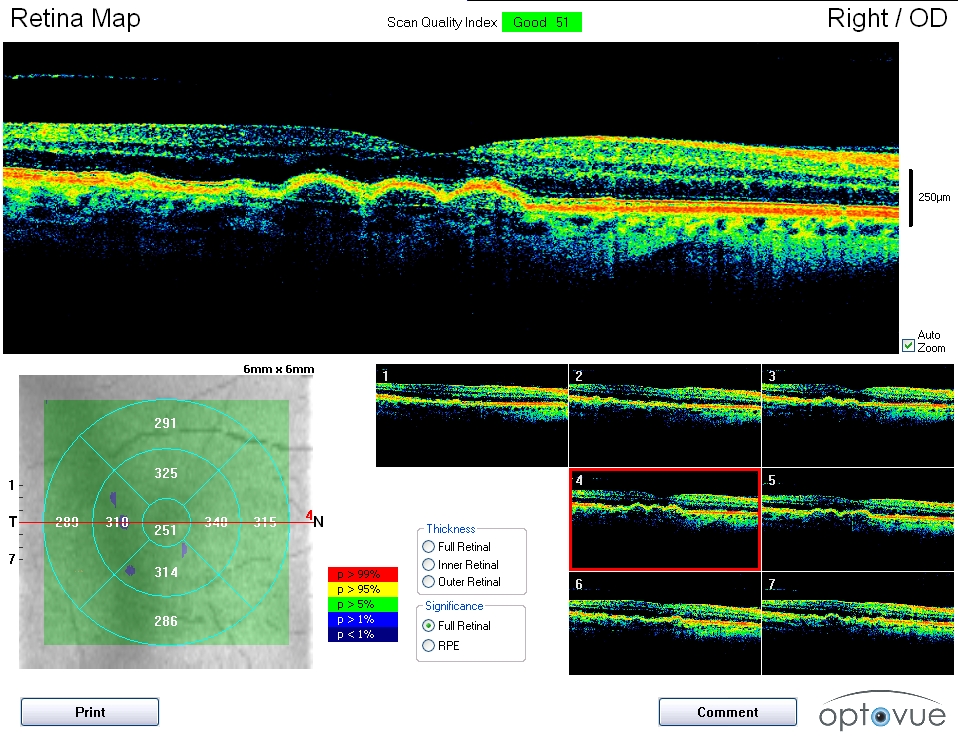
NON EXUDATIVE (DRY) MACULAR DEGENERATION
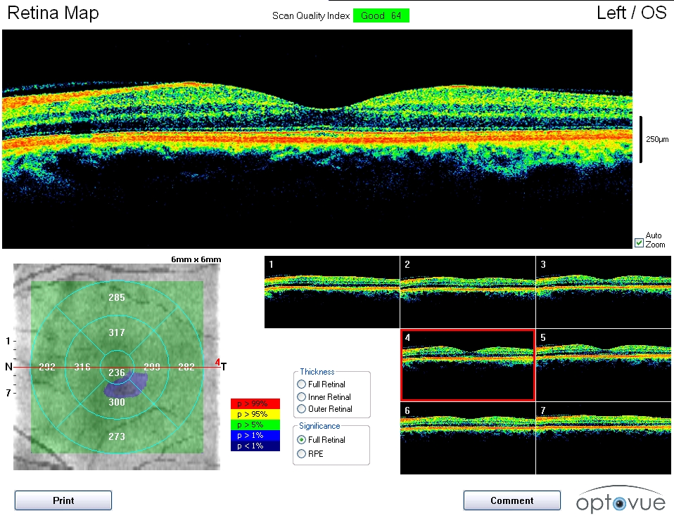
Vitreomacular adhension
Normal cross section of the macula

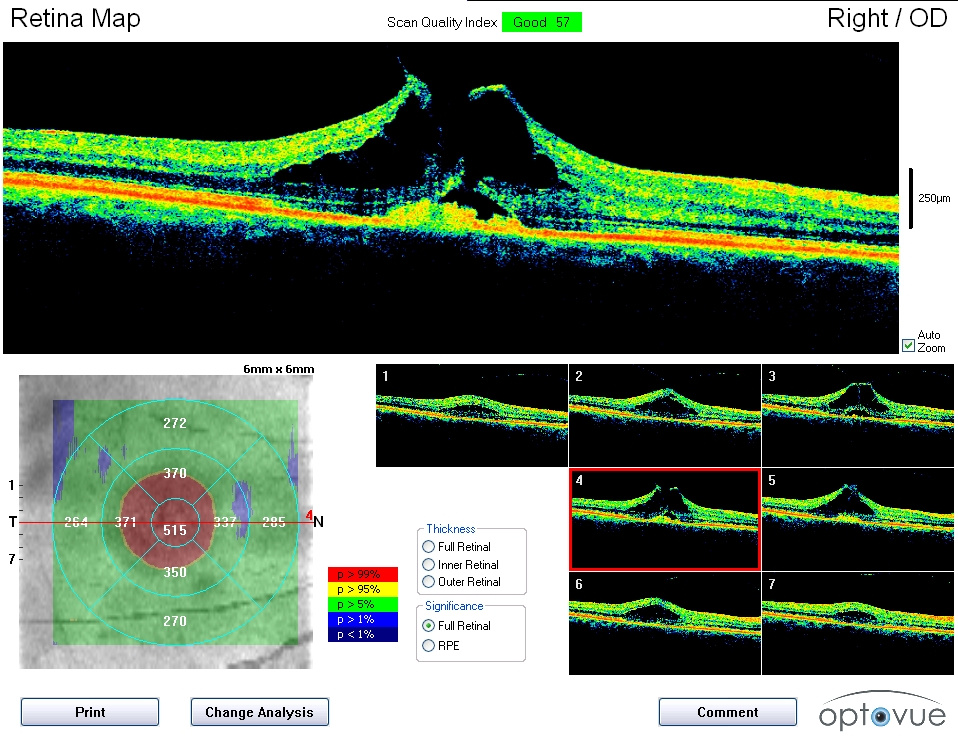
Exudative (Wet) macular degeneration
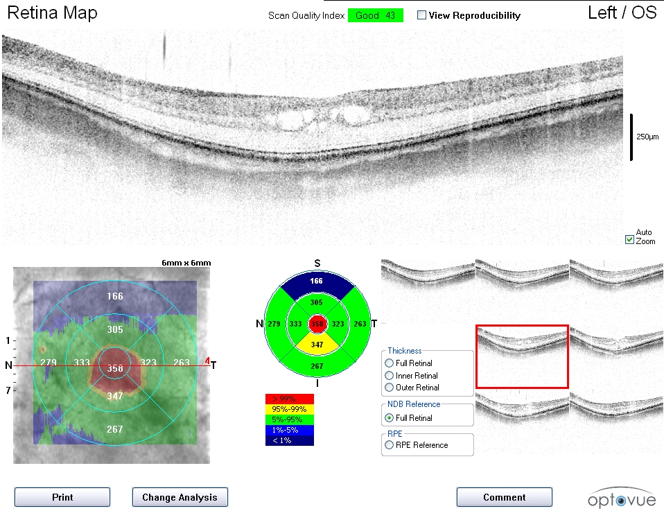
MACULAR EDMA
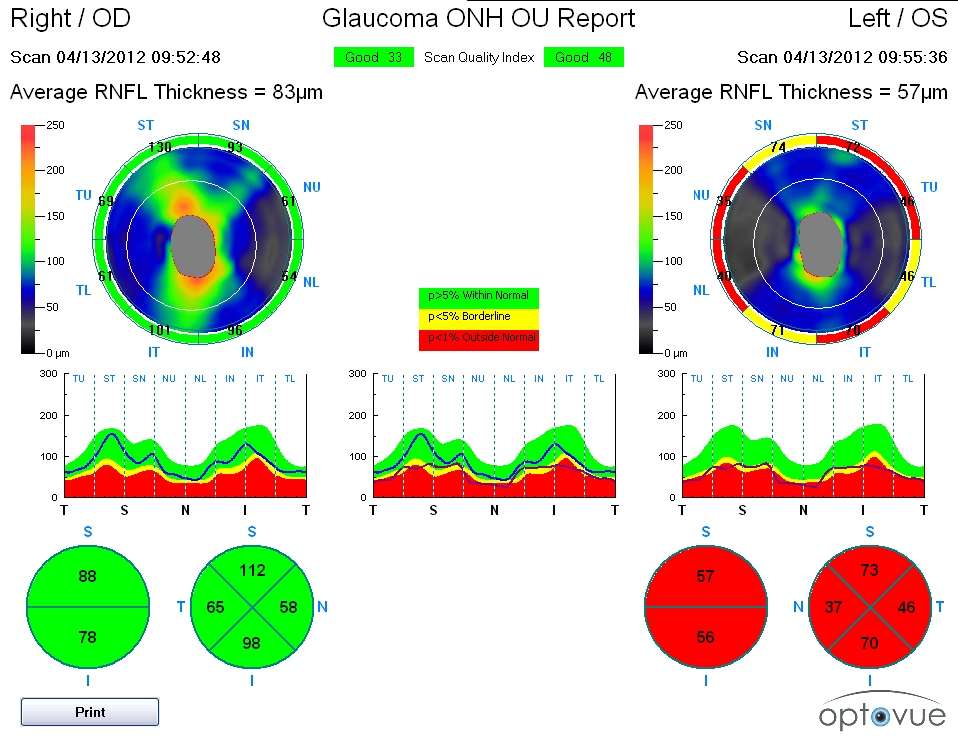
GLAUCOMA